“Francesca, the blood is ready!” I heard from across the room. It was the moment I had been waiting for: my turn to process fresh blood and extract white blood cells to experiment with. I took my lab coat off its hook, put on my gloves, and grabbed my four tubes of Healthy Donor One’s blood. It was time to begin.
This summer I worked in an immunology lab that studies natural killer (NK) cells. NK cells are essential to the function of the immune system. As their name suggests, they are responsible for killing malignant cancer cells or virally infected cells without prior antigen exposure. They migrate through secondary lymphoid tissues, such as the spleen and tonsils. Much like T cells and B cells (two well-known immune lymphocytes), NK cells develop from hematopoietic stem cells (HSCs) and can be extracted from the bloodstream.
This lab studies several different aspects of NK cell function, but my project this summer was focused on migration, which is an essential part of both how cells develop and how they access their target cells. In order to observe and analyze cell migration, cells must be placed under a microscope, and a machine takes various images of the cells to be compiled into a video. With this video, we use computer programs to identify the NK cells and calculate various migration parameters such as distance and speed.
My project compared two different microscopy techniques that the lab uses to study migrating cells: confocal microscopy and in-incubator imaging. Confocal microscopy uses a laser scanner to image cells. The images are high resolution and clearly show the distinct planes that the cells migrate on. It can also create clearer videos because images can be taken within seconds of each other. However, the lasers can be harmful for cells and the process of imaging is very involved. In-incubator imaging takes photos of cells while they are in an incubator at body temperature. It is a less involved process (only requiring the person to press a few buttons), but takes lower-resolution videos with minutes between each frame. Throughout the year, as I write my paper for Fieldston’s science research program, I will quantitatively and qualitatively compare the results from each technique and analyze their pros and cons.
To prepare cells for imaging, it is ideal to use NK cells isolated from fresh blood, and treat them with various cytokines to keep them healthy. The surface of the wells that my cells were in were coated with a ligand that is known to support NK cell migration.
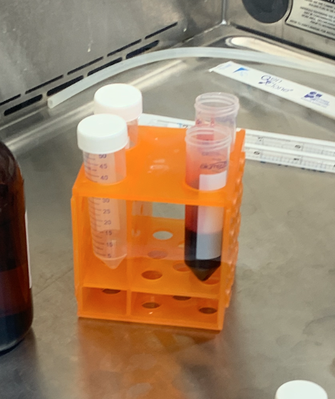
The most memorable part of the internship was certainly the blood processing. On the mornings of experiment days, I would go through a long protocol of extracting NK cells from peripheral blood in a sterile environment. The essential steps of the process were treating the blood with an NK enrichment serum and centrifuging the blood. Centrifuging blood creates layering and separates the red blood cells and plasma. When treated with the NK enrichment serum, a thin, cloudy layer between the platelet-rich plasma and platelet-poor plasma forms that contains the NK cells. This layer is then extracted, the cells are spun in the centrifuge again, and then they are ready for use.

Extracting the NK cell layer.
The cell extraction was the first step to all of my experiments, and it took about two and a half hours to complete. Nonetheless, it was one of my favorite parts of the day. Despite the reddish hue that would develop on my gloves as I poured and pipetted blood from tube to tube, I was thrilled to be touching human tissue and freshly extracting the key ingredient to the lab’s success. Seeing the images and data is the scientific reward for the hands-on activities. As I set up the microscopes and my mentor and I saw cells moving on desktop screens, I knew that every step was worth it.